Credits
Illustrations
The plates on this site are from Henry Gray, Anatomy of the Human Body, 20th edition, edited by Warren H. Lewis (Philadelphia: Lea & Febiger, 1918), drawn largely by Henry Vandyke Carter. They are in the public domain in the US. We use the scans hosted on Wikimedia Commons and recolor them in our navy.
Public domain in the US (published 1918). Each file's Wikimedia Commons license metadata reads "Public domain".
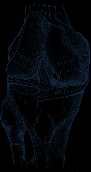
Fig. 347Right knee-joint, from the front, showing interior ligaments. Gray347.png · Wikimedia Commons · Public domain
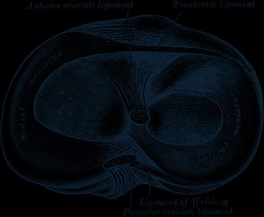
Fig. 349Head of right tibia seen from above, showing menisci and attachments of ligaments. Gray349.png · Wikimedia Commons · Public domain
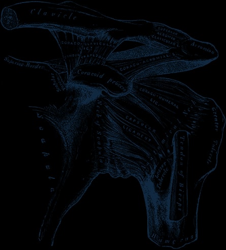
Fig. 326The left shoulder and acromioclavicular joints, and the proper ligaments of the scapula. Gray326.png · Wikimedia Commons · Public domain
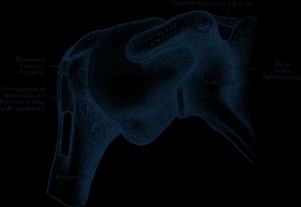
Fig. 327Capsule of shoulder-joint (distended). Anterior aspect. Gray327.png · Wikimedia Commons · Public domain
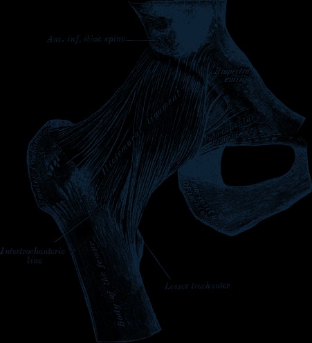
Fig. 339Right hip-joint from the front. Gray339.png · Wikimedia Commons · Public domain
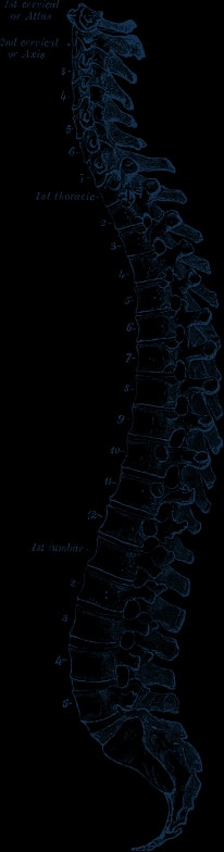
Fig. 111Lateral view of the vertebral column. Gray 111 - Vertebral column.png · Wikimedia Commons · Public domain
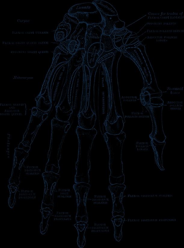
Fig. 219Bones of the left hand. Volar surface. Gray219.png · Wikimedia Commons · Public domain
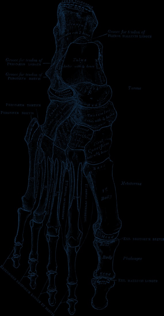
Fig. 268Bones of the right foot. Dorsal surface. Gray268.png · Wikimedia Commons · Public domain
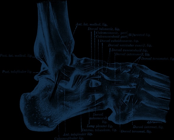
Fig. 355The ligaments of the foot from the lateral aspect. Gray355.png · Wikimedia Commons · Public domain
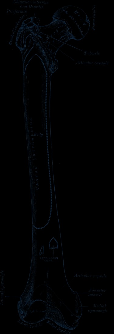
Fig. 244Right femur. Anterior surface. Gray244.png · Wikimedia Commons · Public domain
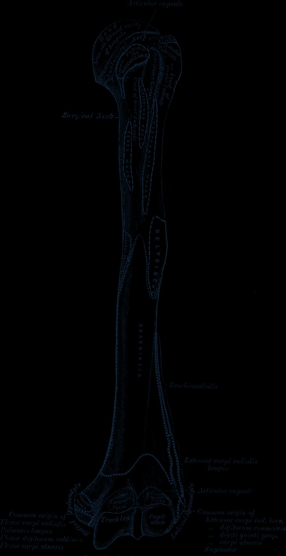
Fig. 207Left humerus. Anterior view. Gray207.png · Wikimedia Commons · Public domain
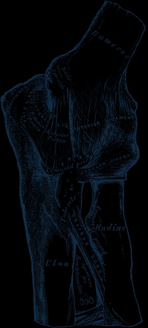
Fig. 329Left elbow-joint, showing anterior and internal ligaments. Gray329.png · Wikimedia Commons · Public domain
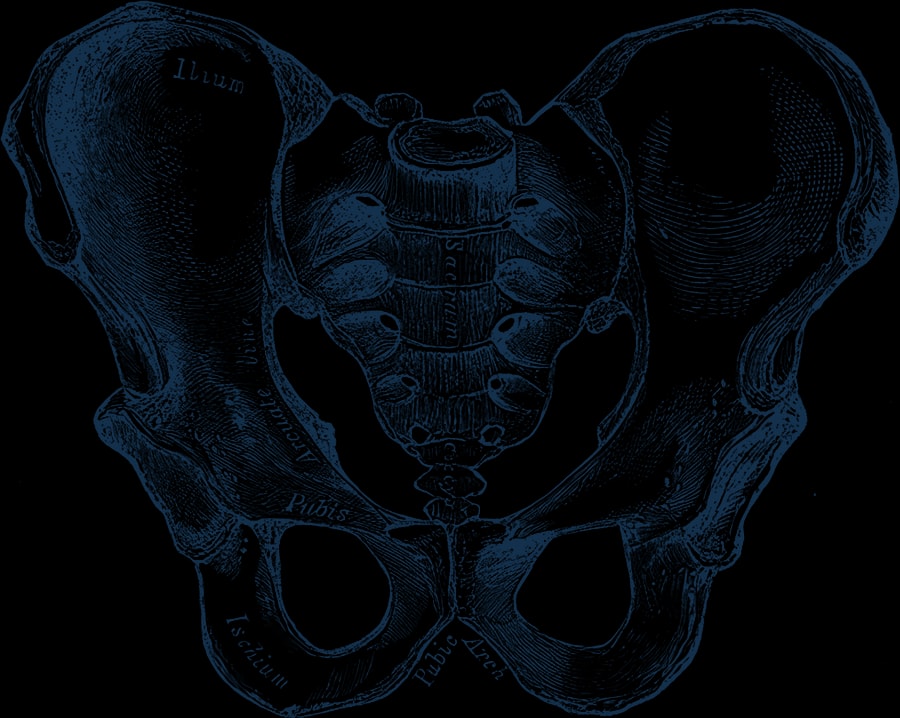
Fig. 241Male pelvis. Gray241.png · Wikimedia Commons · Public domain
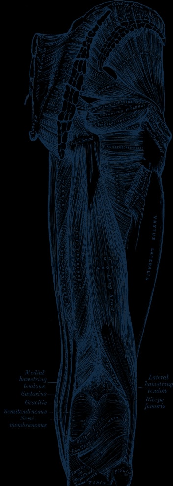
Fig. 434Muscles of the gluteal and posterior femoral regions. Gray434.png · Wikimedia Commons · Public domain